In medicine, X-ray examinations are a fast, reliable and proven method to detect injuries in many specialist areas, e.g. bone fractures and even diseases.
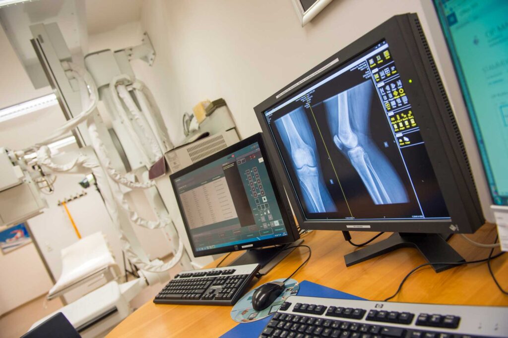
Our X-ray systems are primarily designed for the examination of the skeletal system. At our hospital in Radstadt-Obertauern, we currently work with four digital X-ray machines and two X-ray image intensifiers.
The immediate development of the images in an emergency is particularly important. This enables the easy transmission of the pictures (especially for our guests) by CD or in exceptional cases, by e-mail.
Advantages of the digital X-ray technology
- low radiation exposure
- very good image quality
- possibilities for the post-processing of radiographs
- rapid availability of images for diagnostic purposes
- convenient storage capacity
Fields of application
- In the case of accidents
- Representation of the skeleton
- Representation of the lungs


Procedure of the examination:
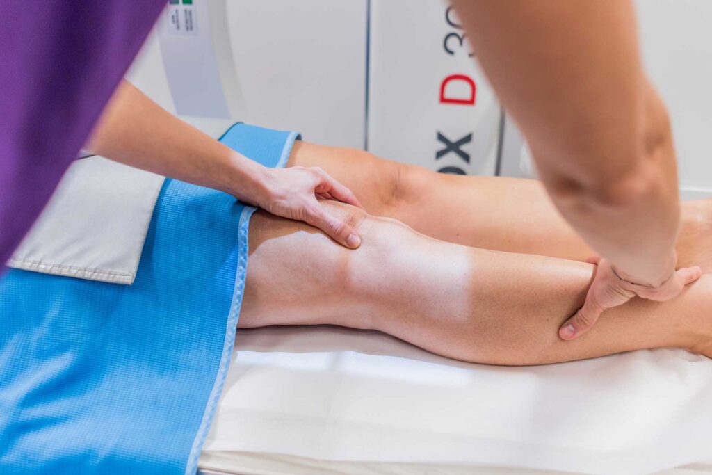
Our digital x-ray machines enable us to produce x-rays of the highest quality with low radiation exposure. During the examination you will be asked to remove clothing and jewellery, depending on the body region to be examined. A few seconds are usually sufficient to create the X-ray image. In order to ensure optimal image quality, it is important that you do not move during the x-ray. The examination is completely painless.
After the images are taken, the x-rays are stored digitally and evaluated. You will also receive your images on CD directly after your examination.


